Malignant Mesothelioma Ct Radiology - The Role Of Imaging In Malignant Pleural Mesothelioma An Update After The 2018 Bts Guidelines Clinical Radiology / The tumor originates from cells of the visceral or parietal pleural and is linked to asbestos exposure with a median latency of 44.6 years .due to the latency between exposure and onset of mesothelioma and the ongoing use of asbestos in parts of the world, the incidence is expected to rise.
Malignant Mesothelioma Ct Radiology - The Role Of Imaging In Malignant Pleural Mesothelioma An Update After The 2018 Bts Guidelines Clinical Radiology / The tumor originates from cells of the visceral or parietal pleural and is linked to asbestos exposure with a median latency of 44.6 years .due to the latency between exposure and onset of mesothelioma and the ongoing use of asbestos in parts of the world, the incidence is expected to rise.
Malignant Mesothelioma Ct Radiology - The Role Of Imaging In Malignant Pleural Mesothelioma An Update After The 2018 Bts Guidelines Clinical Radiology / The tumor originates from cells of the visceral or parietal pleural and is linked to asbestos exposure with a median latency of 44.6 years .due to the latency between exposure and onset of mesothelioma and the ongoing use of asbestos in parts of the world, the incidence is expected to rise.. Khleif, darko pucar radiology and imaging Treatment strategies for mesothelioma often include a combination of surgery and chemotherapy. malignant pleural mesothelioma (mpm) is an aggressive thoracic malignancy with a dismal prognosis. However, tumor invasion into the chest wall or diaphragm can be underestimated by ct. Computed tomography is the primary imaging method used for the diagnosis and the staging of malignant mesothelioma, but also for guiding biopsy for tissue diagnosis.
Primary malignant neoplasm arising from peritoneum. 1 , ,2 2 and and3 3 ). Computed tomography is the primary imaging modality used for the diagnosis and staging of mpm. Wechsler rj, rao vm, steiner rm. ct chest is considered the initial imaging modality for malignant mesothelioma with the limitation of evaluating local invasion and differentiating benign or malignant soft tissue abnormalities.
Double Cancer Comprising Malignant Pleural Mesothelioma And Squamous Cell Carcinoma Of The Lung Treated With Radiotherapy A Case Report from www.spandidos-publications.com Sheets of tumor can be followed inferiorly along the diaphragmatic crura. Role of ct, mri, and pet/ct in staging evaluation and treatment considerations. Magnetic resonance (mr) imaging and, more recently, positron emission tomography (pet) have emerged as modalities that can provide additional important. Thoracic computed tomography findings in malignant mesothelioma. Computed tomography findings in 66 patients with malignant pleural mesothelioma due to environmental exposure to asbestos. Mpm presents with several ct features similar to more common pleural diseases such as metastatic pleural malignancy. Staging of malignant pleural mesothelioma: Rare, mesenchymal primary tumors of the visceral pleura less common than diffuse malignant mesothelioma;
malignant mesothelioma is a rare and aggressive cancer.
Mpm is a rare malignant neoplasm of pleura that represents with several ct features seen in other common pleural malignancies.the aim of this study is to evaluate the agreement of radiology and pathology in differentiation between mpm and metastatic pleural malignancy and to demonstrate most characteristic ct finding of them.material and methods:55 patients,29males and 26females. Pleural biopsies in patients with suspected malignant pleural mesothelioma (mpm) are often inconclusive resulting in repeat diagnostic procedures. Experience of a tertiary referral center It can have a myxoid stroma resulting in a low attenuation on ct and a high attenuation on t2wi. Staging of malignant pleural mesothelioma: (1) right lung, (2) spine, (3) left lung, (4) ribs, (5) descending part of the aorta, (6) spleen, (7) left kidney, (8. We investigated pleural cle imaging as a biopsy guidance technique to distinguish malignant from benign pleural disease. Primary malignant neoplasm arising from peritoneum. Corson n, sensakovic wf, straus c, starkey a, armato sg 3rd. Heelan rt, rusch vw, begg cb, et al. Comparison of ct and mr imaging. 1 , ,2 2 and and3 3 ). Guest pj, reznek rh, selleslag d, et al the first two decades of life 1, 2, and mri is required to define the extent (1992) peritoneal mesothelioma:
547 ct findings of malignant pleural mesothelioma kjronline.org korean j radiol 17(4), jul/aug 2016 diameter of > It can have a myxoid stroma resulting in a low attenuation on ct and a high attenuation on t2wi. The tumors have a distinctive cell type and grow in multiple cysts that are usually benign but in rare cases become malignant. If a doctor suspects you have any form of mesothelioma, benign mesothelioma radiology report or a malignant mesothelioma radiology report, he or she will get your complete medical history and do a physical examination. Role of ct, mri, and pet/ct in staging evaluation and treatment considerations.
Mesothelioma Image Radiopaedia Org from prod-images-static.radiopaedia.org 10 the british thoracic society (bts) statement on malignant mesothelioma specifically recommends scanning at 60 s delay to achieve a. The most common radiologic finding in the ct of patients with malignant peritoneal mesothelioma is ascites. Lftp can be either benign or malignant but is much more often benign (7:1) usually patients are >50 years old 547 ct findings of malignant pleural mesothelioma kjronline.org korean j radiol 17(4), jul/aug 2016 diameter of > The second most common radiologic finding was an infiltration of the small bowel mesentery by tumor reported in 41% of patients 2 . Pleural biopsies in patients with suspected malignant pleural mesothelioma (mpm) are often inconclusive resulting in repeat diagnostic procedures. ct scan of a patient with mesothelioma, coronal section (the section follows the plane that divides the body in a front and a back half). Heelan rt, rusch vw, begg cb, et al.
malignant pleural mesothelioma (mpm) is a rare malignant neoplasm of the pleura that typically affects individuals occupationally exposed to asbestos through a variety of industries.
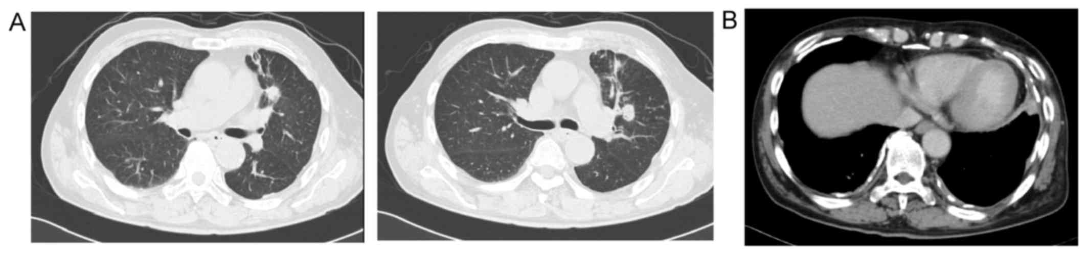
This review is based on a presentation given by angela levy and adapted for the radiology assistant by robin smithuis. It can have a myxoid stroma resulting in a low attenuation on ct and a high attenuation on t2wi. A) axial ct scan, nodular pleural thickening (white arrow), b) sagittal chest ct scan of the soft tissue. ct of malignant pleural mesothelioma. However, here we report a rare case of mpm diagnosed in a healthy young male patient without significant asbestos exposure. Computed tomography of malignant pleural mesothelioma. Corson n, sensakovic wf, straus c, starkey a, armato sg 3rd. Asbestos plaques are not a… While the exact cause of mesothelioma is unknown, the primary risk factor is exposure to asbestos fibers. At ct, the soft tissue density of tumor tissue can be readily distinguished from the adjacent pleural effusion, but the nodules may on occasion be so. Computed tomography (ct) enables early detection of small pleural tumors and pleural effusions and definition of the extension of the tumor along the pleural surfaces and fissures. ct chest is considered the initial imaging modality for malignant mesothelioma with the limitation of evaluating local invasion and differentiating benign or malignant soft tissue abnormalities. For the evaluation of lymph nodes, internal mammary nodes were considered abnormal when
Oncology case 7 june 19, 2019; Mpm presents with several ct features similar to more common pleural diseases such as metastatic pleural malignancy. Asbestos plaques are not a… However, tumor invasion into the chest wall or diaphragm can be underestimated by ct. 547 ct findings of malignant pleural mesothelioma kjronline.org korean j radiol 17(4), jul/aug 2016 diameter of >
Metastatic Malignant Pleural Mesothelioma Masquerading As A Case Of Acute Abdomen Secondary To Small Bowel Perforation Annals Of Saudi Medicine from www.annsaudimed.net Cystic mesothelioma is a very rare form of mesothelioma that originates in the peritoneum of the abdominal cavity. Primary malignant neoplasm arising from peritoneum. We herein report two cases of malignant pleural mesothelioma with marked lymphangiosis. Staging of malignant pleural mesothelioma: Asbestos, chromosomal deletions, and tumor suppressor gene alterations in human malignant mesothelioma. However, tumor invasion into the chest wall or diaphragm can be underestimated by ct. malignant pleural mesothelioma (mpm) is a rare malignant neoplasm of the pleura that typically affects individuals occupationally exposed to asbestos through a variety of industries. In each case the ct showed.
Purpose to identify correlation between ct imaging features and histologic subtypes of malignant peritoneal mesothelioma (mpm).
About cancer cancer research uk. The tumors have a distinctive cell type and grow in multiple cysts that are usually benign but in rare cases become malignant. Okten f, köksal d, onal m, ozcan a, sims¸ek c, ertürk h. 10 the british thoracic society (bts) statement on malignant mesothelioma specifically recommends scanning at 60 s delay to achieve a. Computed tomography of malignant pleural mesothelioma. Role of ct, mri, and pet/ct in staging evaluation and treatment considerations. Mazurek jm, syamlal g, wood jm, hendricks sa, weston a. However, here we report a rare case of mpm diagnosed in a healthy young male patient without significant asbestos exposure. Fdg pet/ct has been increasingly used for the characterization, staging and restaging of mpm recently (figs. Mmwr morb mortal wkly rep. Treatment strategies for mesothelioma often include a combination of surgery and chemotherapy. Average life expectancy for this aggressive cancer is about 12 months after diagnosis. malignant pleural mesothelioma is a rare tumor.
0 Response to "Malignant Mesothelioma Ct Radiology - The Role Of Imaging In Malignant Pleural Mesothelioma An Update After The 2018 Bts Guidelines Clinical Radiology / The tumor originates from cells of the visceral or parietal pleural and is linked to asbestos exposure with a median latency of 44.6 years .due to the latency between exposure and onset of mesothelioma and the ongoing use of asbestos in parts of the world, the incidence is expected to rise."


0 Response to "Malignant Mesothelioma Ct Radiology - The Role Of Imaging In Malignant Pleural Mesothelioma An Update After The 2018 Bts Guidelines Clinical Radiology / The tumor originates from cells of the visceral or parietal pleural and is linked to asbestos exposure with a median latency of 44.6 years .due to the latency between exposure and onset of mesothelioma and the ongoing use of asbestos in parts of the world, the incidence is expected to rise."
Post a Comment